. 2016 Nov 17;5:e21491. doi: 10.7554/eLife.21491

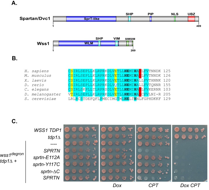
Figure 1. SPRTN fails to complement Wss1 activity in yeast.

(A) Comparison of SPRTN and Wss1 domain organization. (B) Comparison of SprT-like domains from selected SPRTN-containing organisms. Amino acid highlighted in red corresponds to the tyrosine mutated in patients with Ruijs-Aalfs syndrome (SPRTN-Y117C), identical amino acids are in blue and similar amino acids are in yellow. The catalytic glutamic acid and zinc-coordinating histidines are indicated by bold text. (C) Yeast spot assay. Five-fold dilutions of wildtype, tdp1∆, and wss1∆ tdp1∆ cells. wss1∆ tdp1∆ cells harboring empty plasmid (--- = pRS415) or plasmid encoding wild-type SPRTN or corresponding mutants under the endogenous Wss1 promoter. Cells were spotted on CSM-LEU plates with 2 ug/mL doxycycline or 2 ug/mL doxycycline and 40 uM CPT. Doxycycline is used to acutely deplete Wss1 protein levels in tdp1∆ cells to effectively obtain wss1∆ tdp1∆ cells.

DOI: http://dx.doi.org/10.7554/eLife.21491.002

Figure 1.

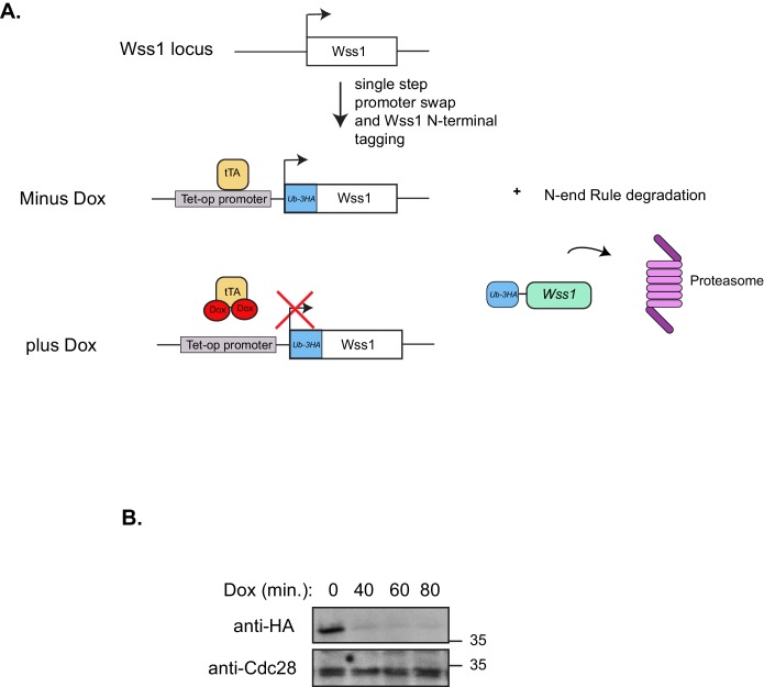
Figure 1—figure supplement 1. Dual Wss1 protein depletion system.

Figure 1—figure supplement 1.

(A) Schematic illustration describing the dual protein depletion system used to acutely deplete Wss1 from tdp1∆ yeast strains to circumvent the accumulation of cells containing second site suppressors that give resistance to CPT. The endogenous Wss1 promoter and the N-terminal region of Wss1 was replaced, in a single step, with a selection cassette that includes a repressible heterologous promoter and ubiquitin-3HA epitope tag. (B) Western blot analysis confirming Wss1 depletion upon doxycycline [2 ug/mL] addition to yeast strain yKM10.
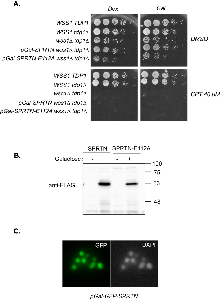
Figure 1—figure supplement 2. SPRTN complementation in yeast.

Figure 1—figure supplement 2.

(A) Galactose inducible expression of FLAG-SPRTN or corresponding mutants integrated at the URA3 locus of wss1∆ tdp1∆ cells. Five-fold serial dilutions of yeast cells were spotted on rich plates containing either 2% glucose with or without 40 uM CPT or 2% galactose with our without 40 uM CPT. (B) Western blot analysis of FLAG-SPRTN and FLAG-SPRTN-E112A expression in yeast cells. (C) Epi-fluorescent microscopy of fixed cells expressing galactose inducible GFP-SPRTN. Cells were stained with DAPI to locate the nucleus.