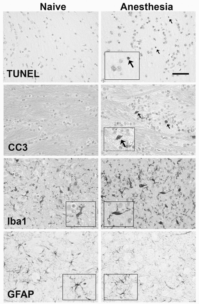
Fig 1. Histology of TUNEL, CCasp-3, Iba-1 and GFAP.
Representative photomicrograpths of TUNEL and CCasp-3 in the external capsule and the Iba-1 and GFAP in the thalamus of naïve (left column) and isoflurane exposed (right column) piglets. Arrows indicate TUNEL and CCasp-3 positive cells. CCasp-3 and Iba-1 are counterstained with cresyl violet. Inserts highlight cell activation and morphology (scale bar = 50μm).

