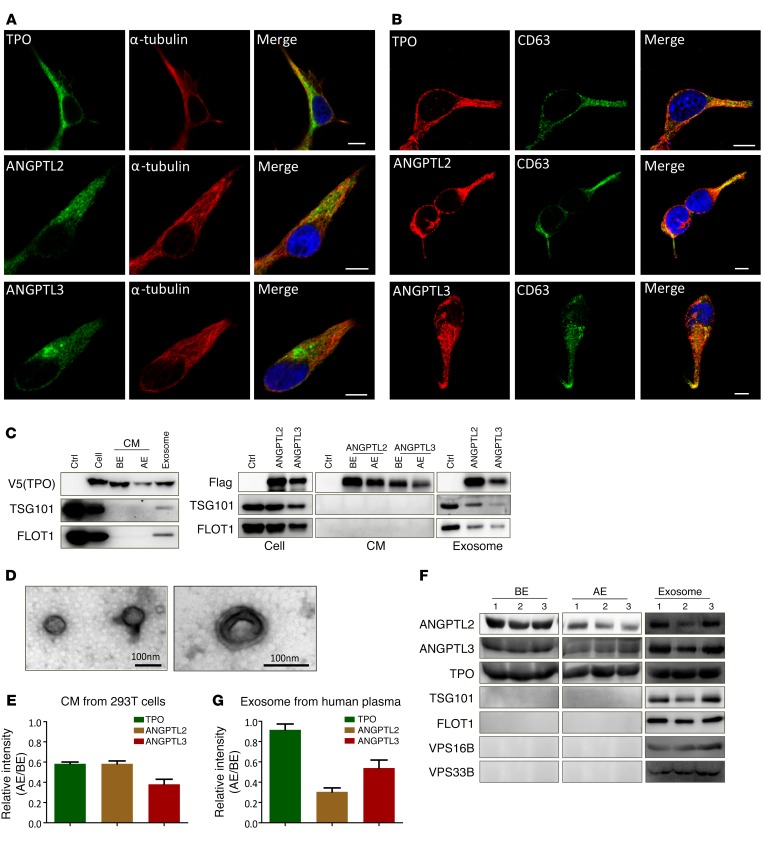
Figure 1. Stemness-related secretory proteins exist in exosomes.
(A and B) 293T cells transfected with TPO, ANGPTL2, and ANGPTL3 were costained with either α-tubulin (A) or CD63 (B) using immunofluorescence staining to evaluate the colocalization, as shown in the merged images of each row. Nuclei were counterstained with DAPI. Scale bars: 10 μm. (C) Exosomes were purified from TPO-, ANGPTL2- and ANGPTL3-conditioned medium (CM) of 293T cells and immunoblotted with antibodies against V5 (for TPO), Flag (for ANGPTL2 and ANGPTL3), and TSG101 and FLOT1 (exosome markers). Ctrl, empty vector; BE, before extraction; AE, after extraction. (D) Representative transmission electron micrographs of exosomes purified from ANGPTL2-conditioned medium of 293T cells (scale bars: 100 nm). (E) Quantitative data for the levels of TPO-, ANGPTL2-, and ANGPTL3-containing exosomes in supernatant after differential centrifugation (AE/BE, n = 3). (F) Exosomes were purified from human plasma and assayed to detect TPO, ANGPTL2, ANGPTL3, TSG101, FLOT1, VPS16B, and VPS33B. (G) Quantitative data for the levels of TPO-, ANGPTL2-, and ANGPTL3-containing exosomes in human plasma after differential centrifugation (AE/BE, n = 3). Experiments were conducted 3 to 5 times for validation.

