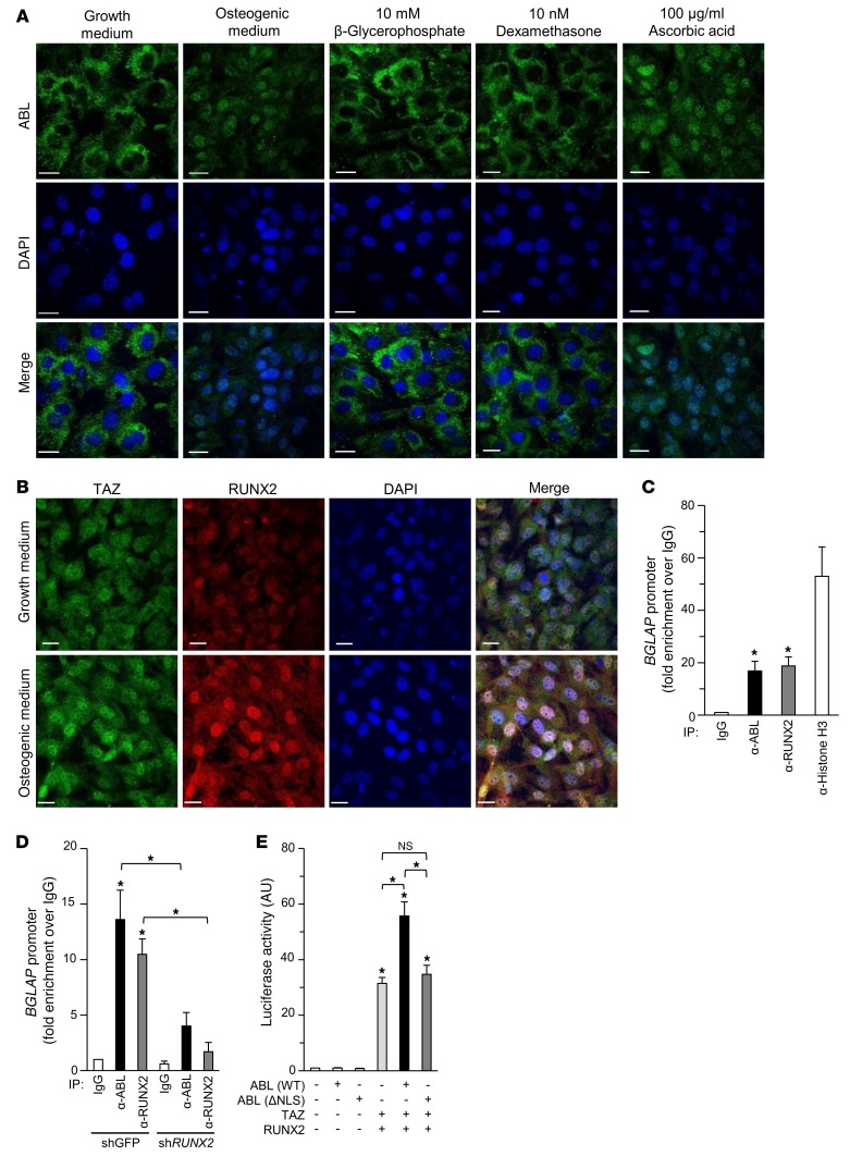
Figure 3. Nuclear ABL is required for the RUNX2-TAZ complex transcriptional activity.
(A) Primary murine osteoblasts were cultured in growth medium, osteogenic medium, or growth medium supplemented with 10 mM β-glycerophosphate, 10 nM dexamethasone, or 100 μg/ml ascorbic acid and stained by immunofluorescence. Scale bars: 20 μm. The images of intracellular ABL (green) and nuclei (blue) are representative of 3 independent experiments. (B) Primary murine osteoblasts were cultured in growth medium or osteogenic medium and stained by immunofluorescence. Scale bars: 20 μm. The images of intracellular TAZ (green), RUNX2 (red), and nuclei (blue) are representative of 3 independent experiments. (C and D) qPCR of chromatin immunoprecipitates from Saos-2 cells (C) or Saos-2 cells infected with shGFP or shRUNX2 (D). Amplicons were designed to flank the RUNX2-binding site within the BGLAP promoter. Fold enrichment represents the signal obtained after IP with a nonspecific IgG antibody. n = 3. (E) Luciferase activity from a BGLAP reporter assay in HEK293T cells cotransfected with the indicated constructs. n = 3. *P < 0.05, by ANOVA with a Tukey-Kramer post-hoc test. Data represent the mean ± SEM.

