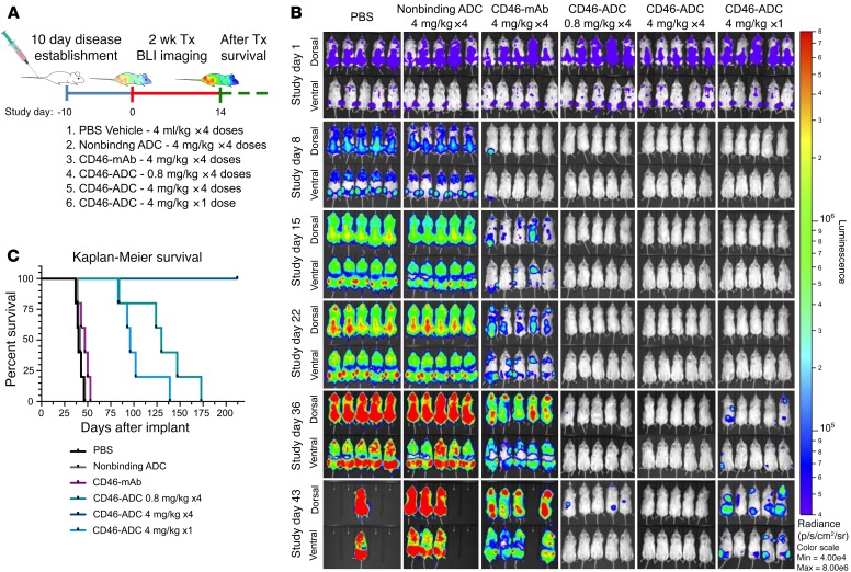
Figure 4. Dose- and schedule-dependent in vivo activity of CD46-ADC in a disseminated MM xenograft model with MM1.S cell line.
(A) Study treatment scheme. MM1.S-Luc cells were injected and established for 10 days. Starting on day 11 (treatment day 1), a total of 4 injections were given twice a week at the concentrations shown for all groups, except for the single-dose group. For each group, n= 5 mice per group. (B) BLI rapidly increased in negative control groups, but decreased to undetectable levels with all CD46-ADC treatment regimens (top views, dorsal; bottom views, ventral). Relapse of disease activity was observed progressively at single-dose 4-mg/kg and low-dose 0.8-mg/kg groups. No detectable BLI signal and no relapse after treatment was observed for the 4-mg/kg, 4-dose schedule, suggesting complete elimination of MM1.S xenografts in vivo. BLI in photons per second per cm2 per steradian (p/s/cm2/sr) was translated to color to indicate disease activity by the legend shown at far right. Tx, treatment; mAb, naked antibody. (C) Kaplan-Meier survival curves of NSG xenografts transplanted with MM1.S-Luc and treated with varying dose levels of CD46-ADC.

