Fig. 3.
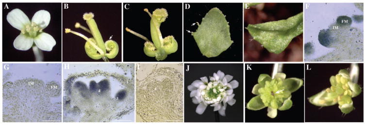
Phenotypes of 35S::MIR172, 35S::AP2, and 35S::AP2m1 lines and in situ localization of miRNA172. (A) A wild-type flower. (B) An ap2-9 flower with first-whorl organs transformed into carpels (arrow). (C) A 35S::MIR172a-1 flower that closely resembles ap2 flowers. A cauline leaf with stigmatic tissues (D, arrows) and a curly rosette leaf (E) from 35S::MIR172a-1 plants. In situ hybridization using an anti-sense [(F) and (H)] or sense [(G) and (I)] miRNA172 probe on longitudinal sections of inflorescence meristems (IM) with two young floral meristems (FM) [(F) and (G)] and stage 7 flowers [(H) and (I)]. Hybridization signals are represented by the blue color. Size bar, 50 μm. The numbers in (H) and (I) represent the positions of the organs, with 1 being the outermost whorl. (J) A 35S::AP2m1 flower with numerous petals and loss of floral determinacy. (K) A 35S::AP2m1 flower with many staminoid organs and an enlarged floral meristem (arrow). (L) A 35S::AP2 flower with more stamens than the wild type.

