
Figure 3.
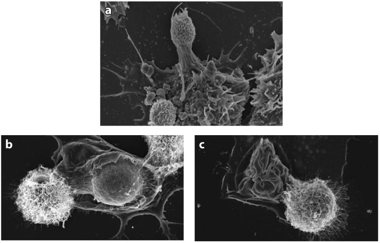
Scanning electron micrographs showing Cryptococcus neoformans and macrophage interaction in vitro. Bone marrow–derived macrophages were infected with antibody-opsonized C. neoformans, and macrophage membranes are shown interacting with yeast cells. (a) Yeast cells are recognized when macrophage membranes probe the extracellular environment around them. (b) Capsulated yeast cells are ingested as the macrophage membrane engulfs them. (c) Ingestion is finalized when the membrane closes upon the yeast cell; a neighboring extracellular yeast is also shown. Panel a courtesy of Sabriya Stukes; panels b and c acquired with the help of Julie M. Wolf.