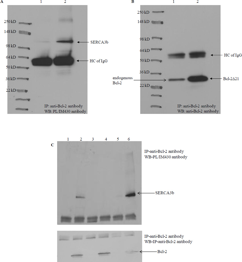
Figure 4. Co-immunoprecipitation of SERCA3b with Bcl-2Δ21 at different SERCA3b:Bcl-2Δ21 molar ratios.
(A) Lane 1: microsomes isolated from SERCA3b overexpressing cells without added Bcl-2Δ21; lane 2: microsomes isolated from SERCA3b overexpressing cells with added Bcl-2Δ21; immunoprecipitation with anti-Bcl-2 antibody and analysis with the PL/IM430 antibody. (B) The membrane used in Figure 4A was stripped and reprobed with the anti-Bcl-2 antibody. Very intense bands around 55 kDa in both A and B might represent the non-specific binding of antibody heavy chains (HC of IgG). (C) Lane 1: microsomes isolated from SERCA3b-encoding vector transfected cells alone; lane 2: microsomes isolated from SERCA3b-encoding vector transfected cells and Bcl-2Δ21 were incubated at a molar ratio of SERCA3b: Bcl-2Δ21 =1:30 at 37°C; lane 3: microsomes isolated from empty vector (without the sequence for SERCA3b) transfected cells; lane 4: microsomes isolated from empty vector (without the sequence for SERCA3b) transfected cells and added Bcl-2Δ21; lane 5: microsomes isolated from SERCA3b overexpressing cells alone; lane 6: microsomes isolated from SERCA3b overexpressing cells and added Bcl-2Δ21 were incubated at a molar ratio of SERCA3b: Bcl-2Δ21=1:6 at 37°C.

