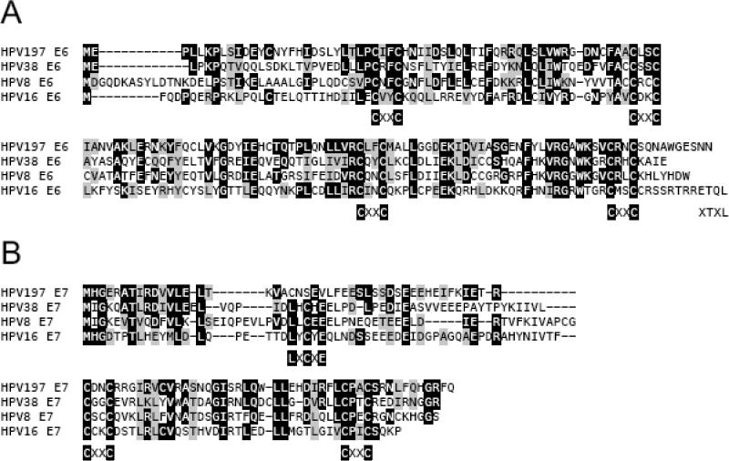
Figure 1.
Sequence alignment of HPV197 E6 (A) and E7 (B) with the corresponding beta 1 HPV8, beta 2 HPV38 and alpha 9 HPV16 proteins. Identical and chemically similar amino acid residues are shown by black and gray boxes, respectively. E7 sequences previously shown to be similar to Adenovirus E1A conserved region (CR) 1 and 2 are shown. The positions of the paired CXXC motifs that form zinc binding sites, the XTXL C terminal PDZ binding site in HPV16 E6 and the LXCXE canonical RB binding site in E7 are also indicated.

