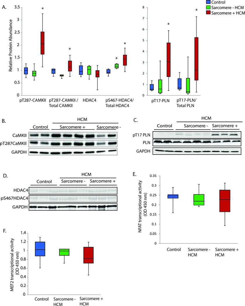
Figure 2.
A. Western blot quantification of autophosphorylated CaMKII (pT287-CaMKII), HDAC4, and the CaMKII phosphorylation targets pS467-HDAC4 and pT17-PLN. Quantification is relative to controls and normalized to GAPDH. For phosphorylation antibodies, quantification is also shown relative to the total antibody quantification. Control n=6, sarcomere mutation positive HCM n=16 (MYBPC3 n=10, MYH7 n=6), sarcomere-negative HCM n=7. B–D. Representative Western blots for CaMKII, HDAC4, and PLN. E–F. NFATc1 and MEF2 transcription factor activity measurement by absorbance at 450 nm wavelength, reference 655 nm. Control n=6, sarcomere mutation positive HCM n=12 (MYBPC3 n=6, MYH7 n=6), sarcomere-negative HCM n=5.. *=p<0.05

