Figure 1.
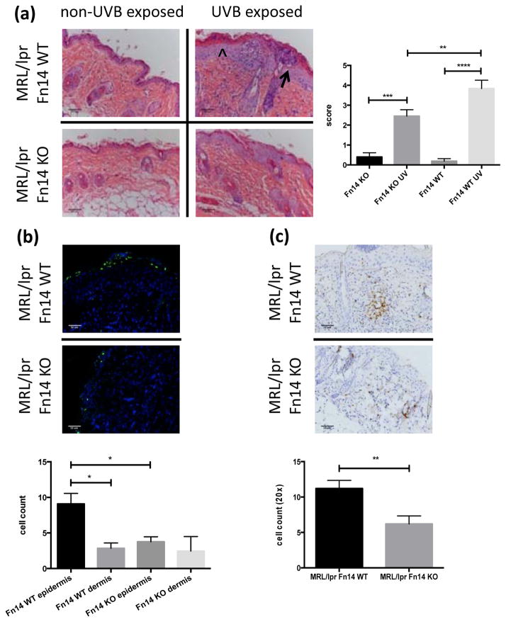
Fn14 deficient MRL/lpr mice are less photosensitive. (a) Shown here are representative H&E images from UVB-exposed and non-exposed skin of MRL/lpr Fn14 WT and KO mice. The arrowhead represents acanthotic epidermis and the arrow points toward an epidermal pustule (left panel). Histopathological skin scores from 13–15 week old female UVB-irradiated MRL/lpr Fn14WT (n=14) and MRL/lpr Fn14KO mice (n=15) are shown in the right panel. (b) Representative images of TUNEL stained paraffin-embedded skin sections from UVB-irradiated MRL/lpr Fn14WT (n=6) and MRL/lpr Fn14KO mice (n=6) (top panel). TUNEL positive cells in the epidermis and dermis were counted using ImageJ (bottom panel). (c) Skin was obtained from UVB-exposed MRL/lpr Fn14 WT and KO mice. Shown are representative images of CD3 stained sections from randomly selected 13–15 week old female UVB-irradiated MRL/lpr Fn14WT (n=7) and MRL/lpr Fn14KO mice (n=7) skin. The bottom panel shows the quantitation of CD3 staining; cells were counted in random 20× fields using ImageJ (WT, n=7; KO, n=7). The figures are representative of 2–3 independent experiments. *P<0.05, **P<0.01, ***P<0.001, ****P<0.0001.

