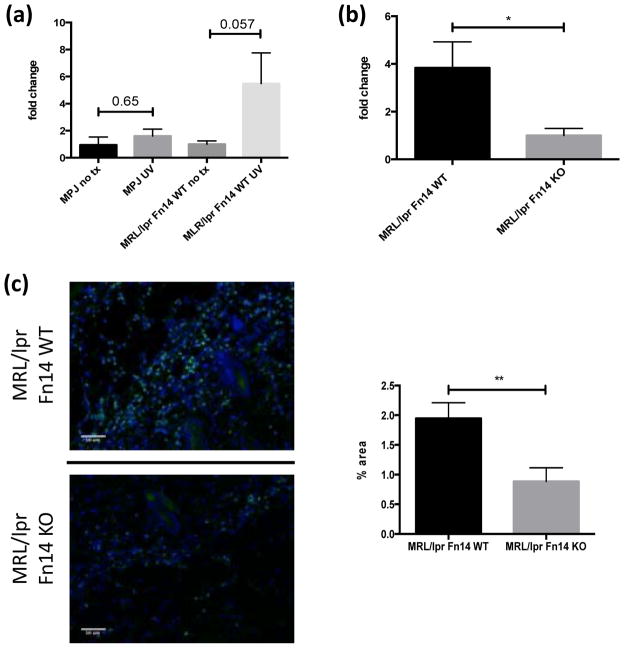
Figure 2.
UVB irradiation induces NGAL. (a) Nine to ten week old female MRL/lpr (n=4) and MRL/MpJ (n=4) mice were irradiated and skin lysates prepared as described above. Depicted in the graph are fold changes in NGAL concentrations compared to baseline, as measured by ELISA. The mean NGAL level in non-irradiated mice in each strain was arbitrarily set at 1. (b) Thirteen to fifteen week old female MRL/lpr Fn14WT (n=8) and Fn14KO (n=7) mice were irradiated, and NGAL in skin lysates analyzed as in (a). (c) Representative images of NGAL stained sections from randomly selected 13–15 week old female UVB-irradiated MRL/lpr Fn14WT (n=8) and Fn14KO mice (n=7). Quantitation of NGAL staining is provided in the right panel. The area with fluorescence was measured by ImageJ. *P<0.05, **P<0.01.

