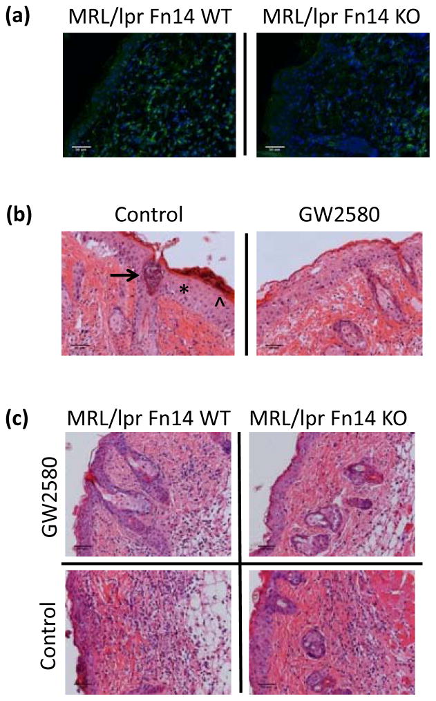
Figure 3.
Macrophage depletion protects MRL/lpr mice from UVB-induced injury. Skin was obtained from UVB-exposed and non-exposed MRL/lpr Fn14 WT and KO mice. (a) Shown are representative images of IBA-1 stained sections from randomly selected 13–15 week old female UVB-irradiated MRL/lpr Fn14WT (n=11) and MRL/lpr Fn14KO mice (n=10). (b) Eight week old female MRL/lpr mice were treated with GW2580 via oral gavage for 16 days. During the last two days of GW2580 treatment, mice were exposed to UVB (50 mJ/cm2) and sacrificed 24 hours later. Shown are representative H&E images from GW (n=5) and PBS-treated (n=6) mice. The asterisk labels acanthotic epidermis, the arrowhead indicates parakeratosis, and the arrow points toward an epidermal pustule (left panel). (c) Representative H&E images of MRL/lpr Fn14WT (GW2580, n=3; PBS, n=2) and MRL/lpr Fn14KO mice (GW2580, n=3; PBS, n=2), treated and irradiated as above.

