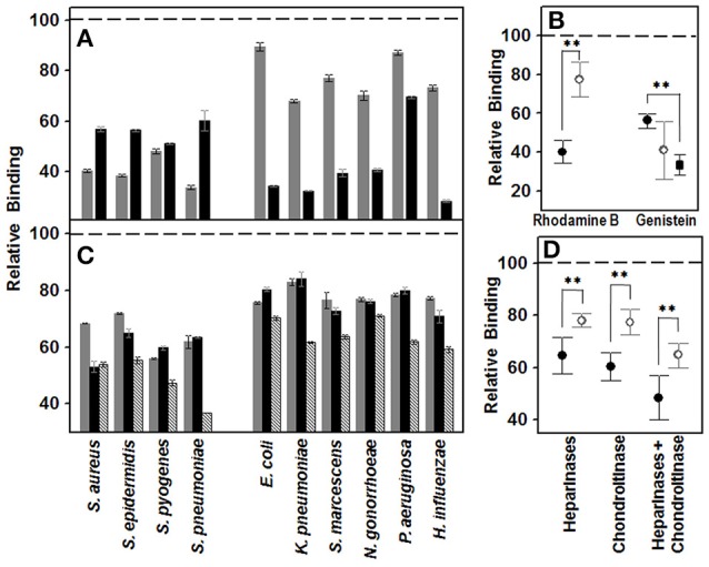
Figure 1.

Effect of decrease in cell GAGs on pathogen adhesion to corneal epithelial cells. (A,B) Effect of inhibition of GAG biosynthesis. (A) Inhibition of bacterial attachment to HCE-2 cells treated with rhodamine B (gray bars) and genistein (black bars). (B) Differences in adherence to HCE-2 cells treated with rhodamine B or genistein between Gram-positive (●) and Gram-negative (○) bacteria. Gram-negative data for binding to genistein-treated cells are represented including or excluding (■) Pseudomonas. (C,D) Effect of the pre-treatment of HCE-2 cell cultures with GAG lyases. (C) Inhibition of bacterial attachment to HCE-2 cells treated with heparinases I and III (gray bars), chondroitinase ABC (black bars) or heparinases + chondroitinase (striped bars). (D) Adherence differences of Gram-positive (●) and Gram-negative (○) bacteria to HCE-2 cells treated with heparinases I and III, chondroitinase ABC or heparinases + chondroitinase. Data were normalized using the adhesion values of bacteria to non-treated cells, which was given the arbitrary value of 1. The spreads represent the standard deviations. Statistically significant differences are denoted by **, which indicates p < 0.01.
