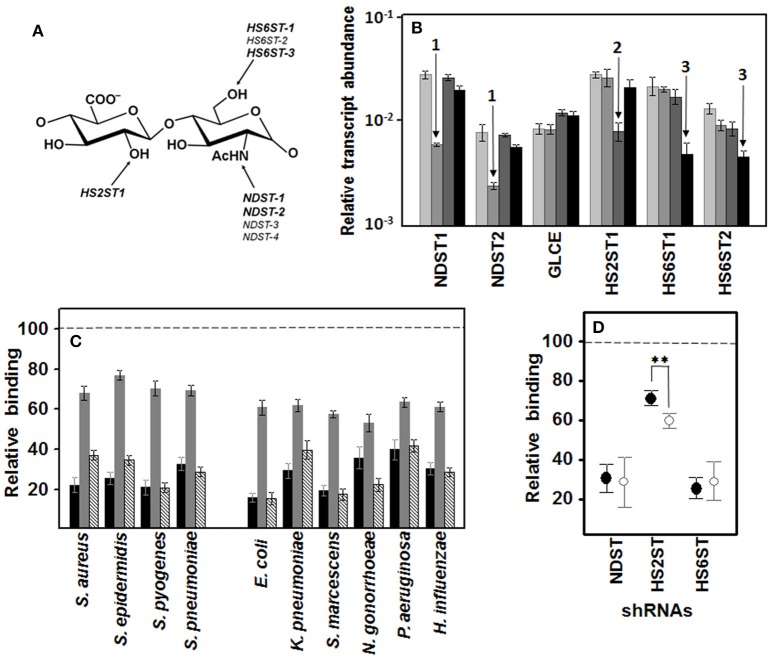
Figure 6.
Influence of specific N- and O- sulfations on pathogen adherence to corneal epithelial cells. (A) Structure of an HS disaccharide unit showing the genes involved in more common sulfations. Genes whose transcription can be detected in corneal epithelial cells are highlighted. (B) Differential transcription of the genes encoding enzymes involved in HS sulfation in HCE-2 cells. The bars with increasingly darker colors indicate the control, and the clones in which N- sulfation, 2-O- sulfation and 6-O-sulfation were silenced, respectively. The arrows indicate downregulation of NDST (1), HS2ST1 (2), and HS6STs (3). NDST1 and -2: N-deacetylase/N-sulfotransferase 1 and 2; GLCE: C5-glucuronate epimerase; HS2ST1: HS 2-O-sulfotransferase; HS6ST1 and -2: HS 6-O-sulfotransferase 1 and 2. (C) Inhibition of bacterial attachment to HCE-2 cells with silencing of the genes involved in N-sulfation (black bars), 2-O- sulfation (gray bars), and 6-O-sulfation (striped bars). (D) Relative adherence differences of Gram-positive (●) and Gram-negative (○) bacteria to clones in which N-sulfation, 2-O-sulfation and 6-O-sulfation were silenced. The spreads represent the standard deviations. Statistically significant differences are denoted by **, which indicates p < 0.01. We used RNA interference to silence the expression of the genes involved in these reactions.

