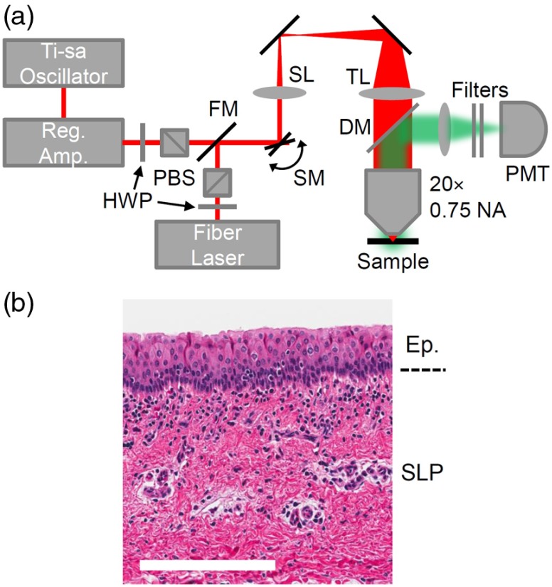
Fig. 2.
Experimental details. (a) Ablation and nonlinear imaging setup including half-wave plates (HWP), polarizing beamsplitters (PBS), a flip mirror (FM), galvanometric scanning mirrors (SM), a scan lens (SL), a tube lens (TL), and an objective. Light emitted from the sample is epicollected by a dichroic mirror (DM) and a photomultiplier tube (PMT). (b) A histological section of a porcine vocal fold, showing the epithelium (Ep.) consisting of tightly packed cells and superior lamina propia (SLP), which is rich in collagen. .

