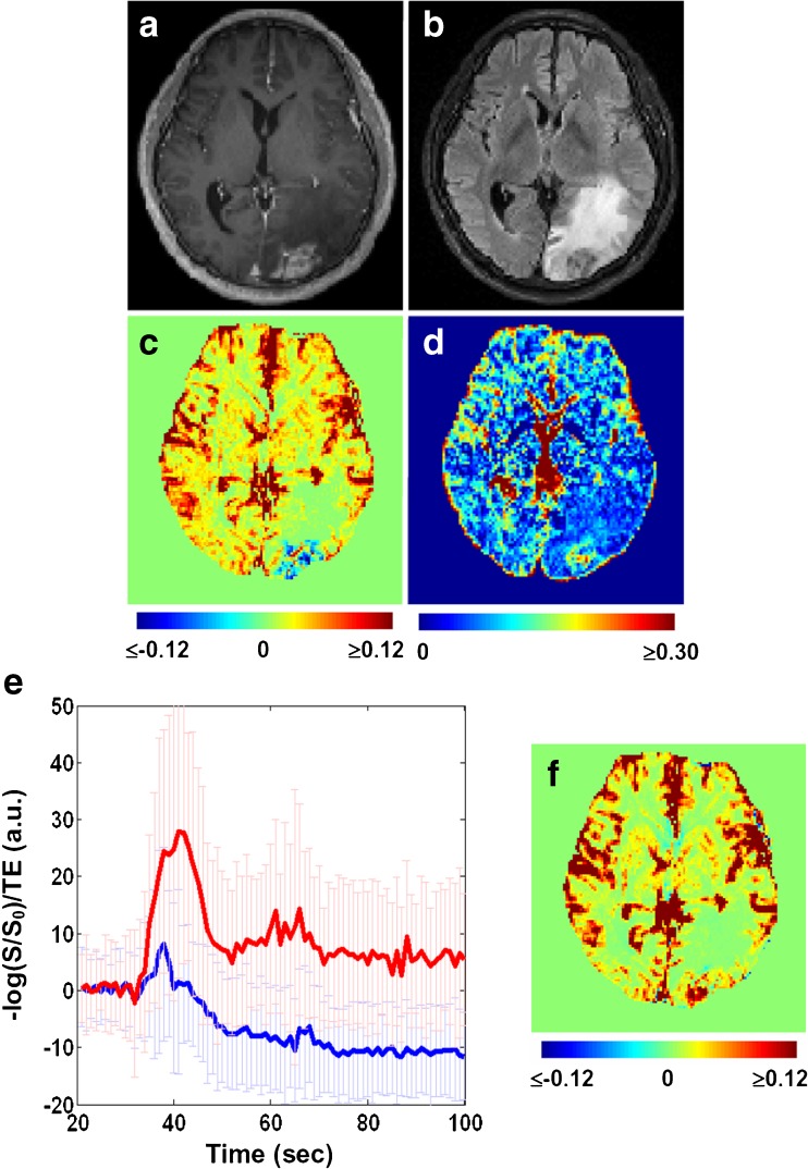
Fig. 3.
Cerebral blood volume derived from dynamic susceptibility contrast imaging (CBVDSC) can be negative in the contrast-enhanced tumour if not corrected for contrast leakage. (a) Post-contrast T1-weighted image. (b) Fluid-attenuated inversion recovery image. (c) Uncorrected CBVDSC map. (d) Map of blood volume fraction f. (e) Concentration time curves extracted from the lesion voxels enhanced in (a) and surrounded by oedema in (b). Amongst these voxels, the ones with negative CBVDSC are plotted in blue while the rest are plotted in red. Error bars indicate the standard deviation across voxels. (f) Corrected CBVDSC map

