
Fig. 4.
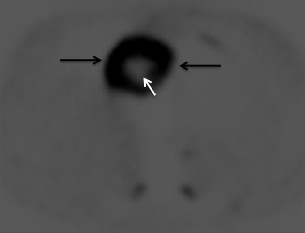
Axial image of (18)F-fluorodeoxyglucose positron emission tomography shows an intensely hypermetabolic lesion in the right heart (black arrows) with central photopenia suggestive of necrosis (white arrow).
Official websites use .gov
A
.gov website belongs to an official
government organization in the United States.
Secure .gov websites use HTTPS
A lock (
) or https:// means you've safely
connected to the .gov website. Share sensitive
information only on official, secure websites.
Axial image of (18)F-fluorodeoxyglucose positron emission tomography shows an intensely hypermetabolic lesion in the right heart (black arrows) with central photopenia suggestive of necrosis (white arrow).