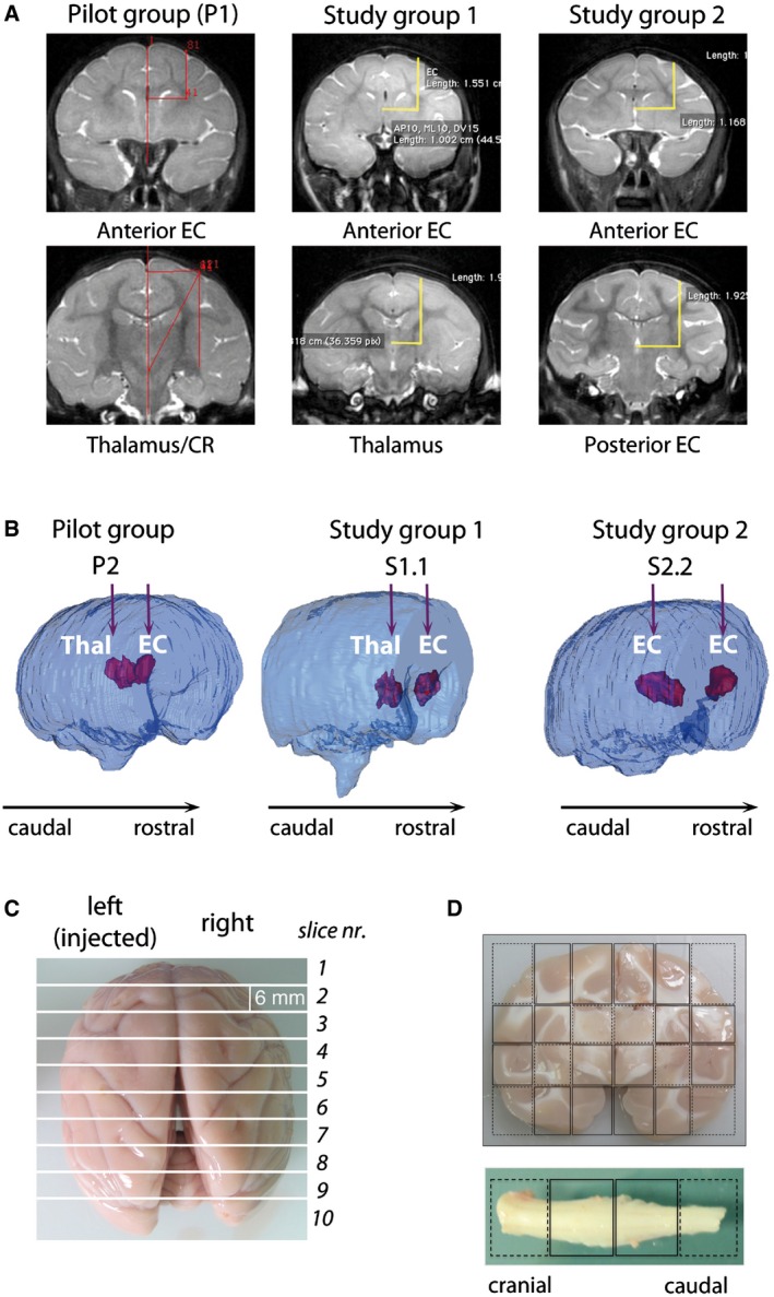
Figure 1. LV.GFP‐ and LV.hARSA‐injected NHP: treatment groups and tissue collection.

- Coordinates for injection in normal NHP of the pilot group, study group 1 and study group 2, were calculated on the bases of pre‐surgery MRI scans.
- 3D rendering of injected brains generated based on post‐surgery MRI scans showing the injection sites and the brain volume containing the vector suspension in the different treatments groups. EC, external capsule; Thal, thalamus; CR, corona radiata; ant, anterior; post, posterior.
- The brain was cut in 6‐mm‐thick slices using an adjustable brain matrix, obtaining 9–10 slices/brain.
- Each brain slice was divided along the midline and each part subdivided into blocks. The cervical spinal cord was cut in four blocks.
