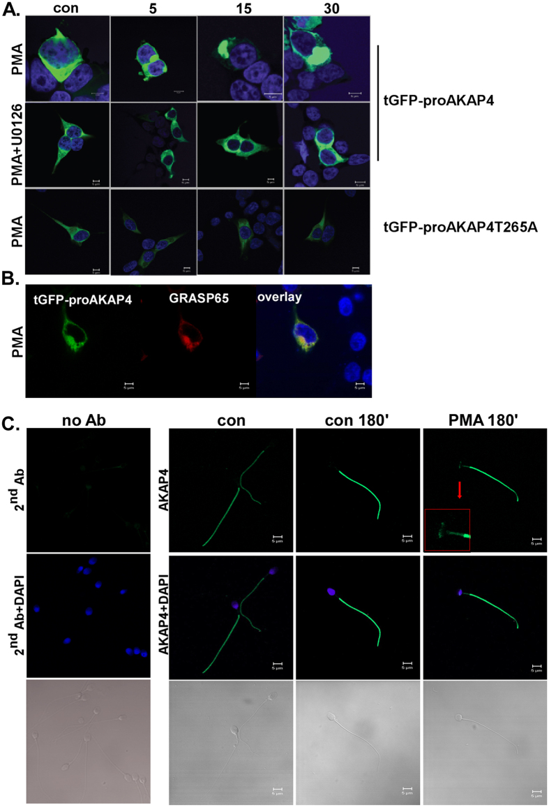
Figure 7. Localization of AKAP4.
(A,B) Fate of AKAP4-tGFP and AKAP4-T265A in HEK293T. HEK293T were transfected with tGFP-proAKAP4 or with tGFP-proAKAP4-T265A. Serum-starved cells, 48 h after transfection, were pretreated with or without U0126 (10 μM) for 20 min. Thereafter PMA (25 nM) was added for different time points. (B) Golgi localization was visualized by cotransfection with GRASP65-RFP. Formalin-fixed slides were imaged under a 63 objective on Zeiss confocal microscope. At least 10 images from each treatment were collected and a representative image is shown. The scale bar is 5 μm. (C) Fluorescence microscopy for expression of AKAP4 in human spermatozoa. Human spermatozoa were treated with PMA (25 nM) for 180 min and then were reacted with anti-AKAP4 antibody and DAPI. Negative control with secondary antibody is shown on the left. Note that AKAP4 is localized to the principal piece under basal conditions and in the principal piece, mid-piece and the post-acrosomal region under PMA stimulation. At least 10 images from each treatment were collected and a representative image is shown. Scale bars indicate 5 μm.

