Figure 5.
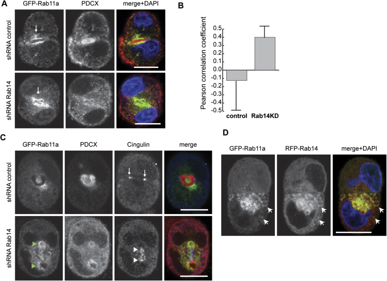
(A) Rab14 KD does not impact the distribution of GFP-Rab11a. Cells over-expressing GFP-Rab11 were labeled for podocalyxin (red). GFP-Rab11a surrounds the apical lumen in both control and Rab14 KD pairs (arrows) and is present in cytoplasmic puncta. However, Rab14 KD results increased cytoplasmic podocalyxin (middle panel). (B) Pearson’s correlation coefficient analysis of peripheral GFP-Rab11a and podocalyxin colocalization. No correlation was observed in control cells. However, Rab14 KD results in an increase in the Pearson’s correlation of endosomal podocalyxin and GFP-Rab11a. (C) Cell pairs over-expressing GFP-Rab11a were co-labeled with cingulin (blue) and podocalyxin (red). In control pairs, cingulin localized to tight junctions (arrows) while GFP-Rab11a surrounds the AMIS. In Rab14 KD pairs, GFP-Rab11 (green arrowheads) accumulates near cingulin (white arrowheads) but there is decreased AMIS-associated podocalyxin. (D) Cells co-transfected with GFP-Rab11a and RFP-Rab14. GFP-Rab11a and RFP-Rab14 colocalize near the AMIS and in the periphery (arrows). Scale bars, 10 μm. DAPI, nuclei (blue).

