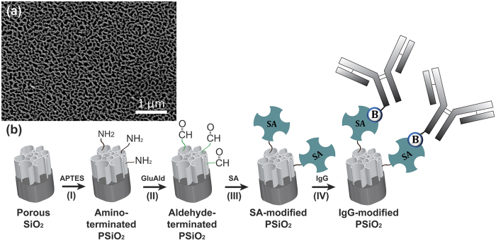
Figure 3.
(a) A top-view high-resolution scanning electron microscope (HRSEM) image of a typical PSiO2 film demonstrating the porous nanostructure morphology with typical pores in the range of 60–100 nm. (b) Schematic illustration of the synthesis steps for the biofunctionalizion of PSiO2 with IgG. (I) PSiO2 was reacted with APTES and catalyzed by an organic base to create an amine-terminated surface. (II) The amine-terminated PSiO2 was reacted with one of the aldehyde groups of the cross-linker GluAld. (III) Grafting of SA onto the aldehyde-terminated surface. (IV) Biotinylated-IgG (E. coli) was conjugated via biotin-SA binding.

