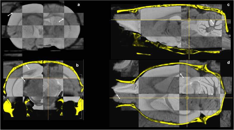
Figure 4. Image-to-template registration.
Checkerboard visualization of corresponding coronal (a,b), sagittal (c) and horizontal (d) cross-sections of the ex vivo MRH atlas template32 and the spatially aligned and resampled in vivo post-operative MR image of a representative animal in our study, with the skull derived from its pre-operative CT overlaid (in yellow, panels b–d). Registration quality can be visually appreciated from the smooth transition between both images at tissue boundaries in different anatomical regions as indicated by the arrows (brain contour, corpus callosum, anterior commissure, white matter fissures of cerebellum).

