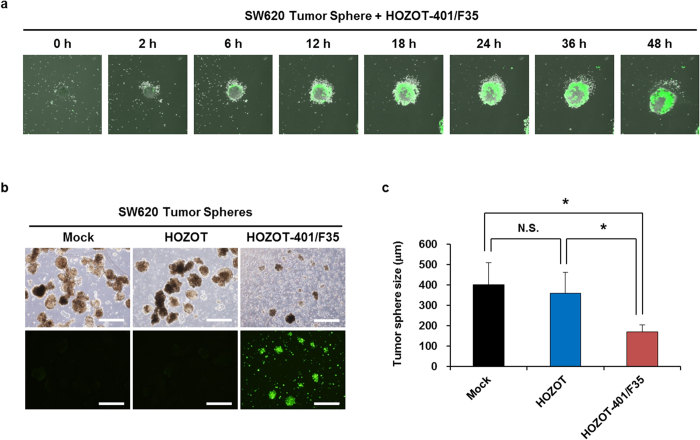
Figure 5. Suppression of SW620 tumor sphere formation by treatment with virus-loaded HOZOT-401/F35 cells.
(a) Time-lapse images of tumor spheres treated with virus-loaded HOZOT-401/F35 cells. (b) Phase-contrast and fluorescence images of tumor spheres from SW620 cells after treatment with mock, virus-free HOZOT cells, or virus-loaded HOZOT-401/F35 cells (left). Scale bars: 500 μm. Tumor size (diameter) was calculated for each group of tumor spheres treated with mock (n = 29), virus-free HOZOT cells (n = 30), or virus-loaded HOZOT-401/F35 cells (n = 18) (right). Data are expressed as mean values ± SD. *P < 0.05. N.S., not significant.

