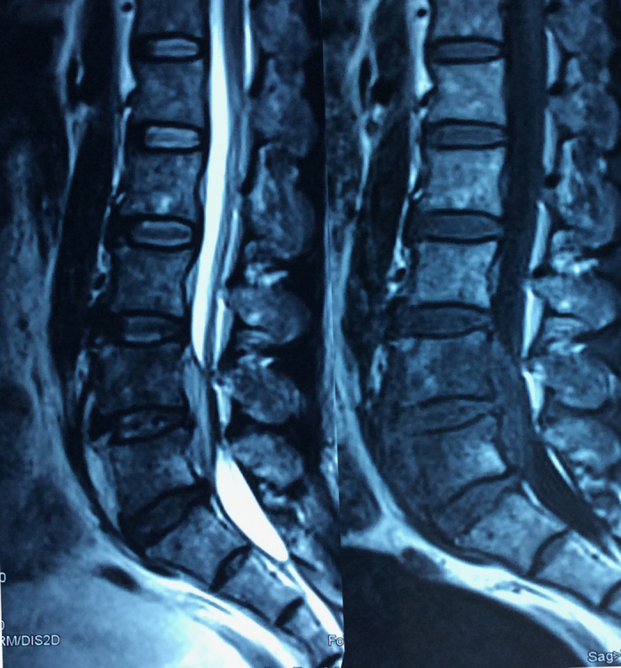
Figure 3.

MRI demonstrated L4–5 spondylodiscitis and epidural abscesses extending to prevertebral region and epidural space causing cauda equina compression. The T1-weighted images of epidural abscesses were hypointense signals, and the signals in these areas became hyperintense on T2-weighted sequences.
