
Figure 2.
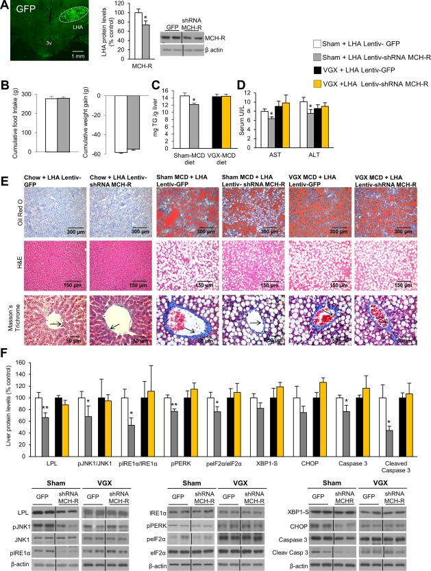
Genetic down‐regulation of MCH‐R in the LHA ameliorates MCD diet–induced NASH and ER stress through the vagus nerve. Representative photomicrographs of brain section showing the injection of a lentivirus that encodes GFP precisely placed in the LHA (×1.25 magnification) and MCH‐R protein levels in the LHA 3 weeks after lentiviral injection encoding either GFP or shRNA MCH‐R (A). Food intake and body weight (B); TG liver content (C); serum levels of AST and ALT (D); representative photomicrograph of liver sections with oil red O, hematoxylin and eosin, and Masson's trichrome staining (E); and liver protein levels of LPL, pJNK, JNK, pIRE1α, IRE1α, pPERK, peIF2α, eIF2α, XBP1S, CHOP, caspase 3, and cleaved caspase 3 (F) in rats fed the MCD diet infected with a lentivirus encoding GFP or shRNA MCH‐R in the LHA in combination with either sham (controls) or vagotomy. Protein β‐actin levels were used to normalize protein levels. Dividing lines indicate splicings within the same gel. Separated photos indicate that gels were run independently. Values are mean ± standard error of the mean of seven or eight animals per group. * P < 0.05, ** P < 0.01, *** P < 0.001 versus controls. Abbreviations: H&E, hematoxylin and eosin; 3v, third ventricle; VGX, vagotomy.