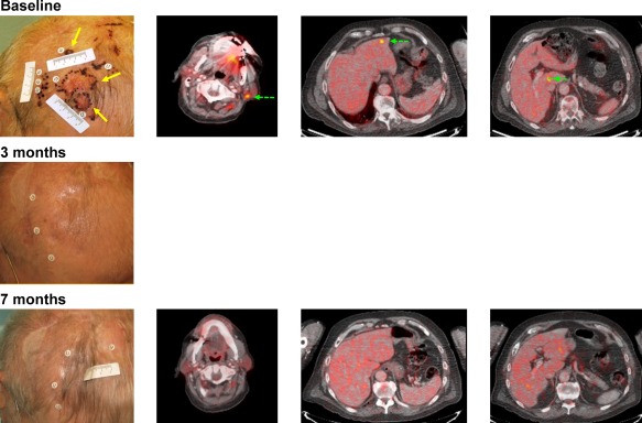
Figure 1.

(A) Representative images from a patient with melanoma of the scalp with metastasis to cervical lymph nodes and liver (stage IVM1c). The patient was diagnosed 2 years before enrollment in OPTiM and had 2 surgeries: one at diagnosis, and another 1 year after recurrence. Top row: injection sites shown in yellow arrows at baseline (left panel). Uninjected sites are shown with green dashed arrows. Black dots mark tumor lesions. Sites included 1 fluorodeoxyglucose (FDG)‐avid left upper level V cervical lymph node (center left panel) and 2 FDG‐avid liver lesions (center right and right panels). Middle row: injections were stopped after complete resolution of scalp lesions after cycle 2 (1 cycle = 2 injections of talimogene laherparepvec). Bottom row: Complete resolution of cervical and liver tumors was documented by FDG‐PET CT at cycle 7. Patient was in complete response until the end of the trial, duration of response (complete response) was approximately 17 months.
