
Figure 1.
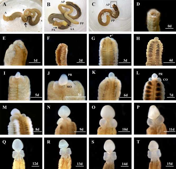
Regenerating anterior structures on Ptychodera flava. A: An intact, live animal. Anterior is to the left. The arrow indicates where the animal will be bisected. a = anterior, p = posterior. B: A bisected animal. AA, anterior end of the anterior half; PA, posterior end of the anterior half; AP, anterior end of the posterior half; PP, posterior end of the posterior half. C: The posterior half of the animal. The boxed area is the site of regeneration. D: The cut site of the posterior half of the animal. E–H: One day (1d) through 4 days (4d) postcut, showing the open wound has healed and a regeneration blastema has formed in two different animals. The arrow in (G) marks the blastema. I,J: At 5 days post bisection (5d), the blastema has formed a rudimentary proboscis (PR) and the mouth (MO) is open on the ventral side. K–T: Six days (6d) through 15 days (15d) show the proboscis (PR) the collar (CO) regenerating around the proboscis stalk in a ventral to dorsal manner. All views are dorsal and anterior is to the top except I, J, which are ventral views. Scale bars = 1 mm.