
Figure 4.
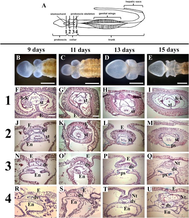
Cross‐sections of Ptychodera flava anterior regeneration days 9–15. All sections are stained with Milligan's trichrome at four different stages of regeneration: 9 days, 11 days, 13 days, and 15 days postamputation. A: Diagram of P. flava showing approximate locations of sections. Row (1) is taken through the posterior proboscis in all four regeneration stages. Row (2) is taken through the anterior collar. Row (3) is taken through the mid collar. Row (4) is taken through the posterior collar. dv, dorsal vessel; E, ectoderm; En, endoderm; h‐k, heart‐kidney complex; Nt, neural tube; ps, proboscis skeleton; st, stomochord. Scale bars = 1 mm in B–E; 0.1 mm in F–U. B–U: Dorsal views with anterior to the right in (B–E) and dorsal to the top in (F–U). All sections are stained with Milligan's trichrome stain. Collagen, green; nuclei, muscle = magenta.