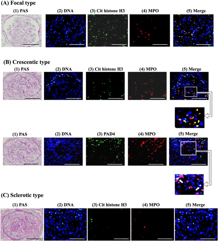
Figure 2.

Representative photomicrographs of histology and neutrophil extracellular trap (NET) staining in the kidney Renal biopsy specimens from 30 patients with myeloperoxidase‐ANCA‐associated microscopic polyangiitis (MPO‐AAV) were examined by periodic acid‐Schiff staining and double immunofluorescences taining for NETs. (A) focal type of necrotizing GN, (B) crescentic type of GN, and (C) sclerotic type of GN. (1) Periodic acid‐Schiff‐staining in A, B, and C (×20), NET staining in A, B and C (2) DNA was stained as blue with Hoechst 33342. Scale bars indicate 100 µm. In A, (3) citrullinated histone H3 is depicted in green, (4) MPO in red, (5) merged images of (2–4). Solid arrows indicate merging signals. In B, on the upper panels (3) citrullinated histone H3 in green, (4) MPO in red, (5) merged images of 2–4. On the lower panels (3) PAD4 in green, (4) MPO in red, (5) merged images of 2–4. Square lines with arrow indicate higher magnification insets. In C, (3) citrullinated histone H3 in green, (4) MPO in red, (5) merged images of 2–4. Yellow indicates the colocalization of citrullinated histone H3 and MPO, PAD4 and citrullinated histone H3.
