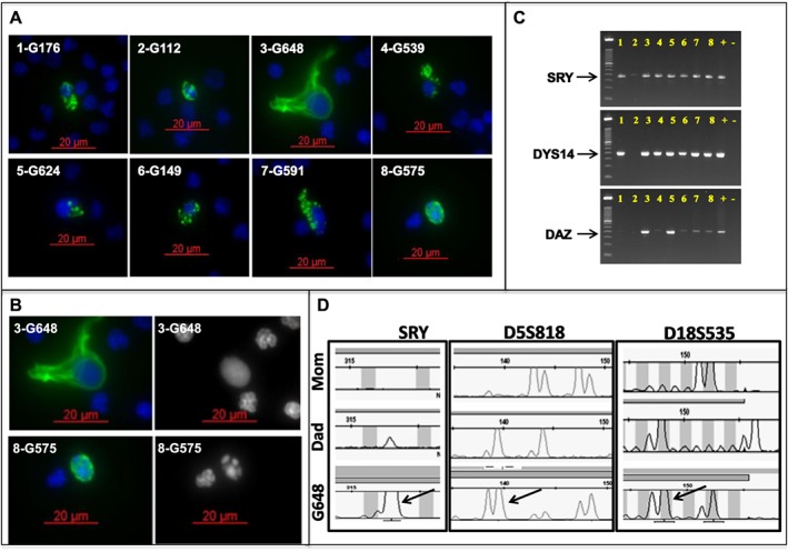
Figure 1.

Multiple fetal trophoblastic cells isolated from one patient. A. Eight fetal cells (confirmed by STR analysis) from a single patient (subject 365) showing the range of nuclear (DAPI; blue) and cytokeratin (CK; green) staining morphology. B. Two of the fetal cells from subject 365 demonstrating uniform (top right) and fragmented (bottom right) DAPI staining. C. Hemi‐nested Y‐chromosome specific PCR performed on the eight fetal cells isolated from subject 365 (male fetus), showing positive amplification of one or more targeted regions on the Y chromosome (SRY, DYS14, DAZ). Numbers correspond to the number prefix for each individual cell in A. D. Ampli1 STR analysis comparing WGA product from a single fetal cell (G648, subject 365) and genomic DNA from each parent. The three loci shown here demonstrate the expected paternal inheritance (arrows) from the Y chromosome (SRY; left panel) and bi‐parental inheritance from chromosomes 5 (D5S818; center panel) and 18 (D18S535; right panel)
