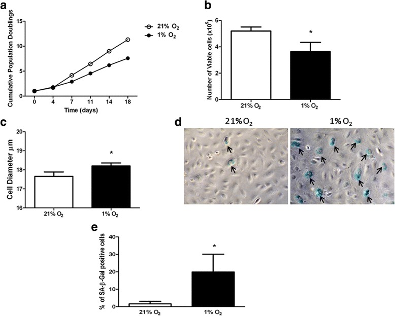
Fig. 4.

Long-term exposure of ECFCs to hypoxia induces partial senescence. a ECFCs cultured at 21% O2 (open circles) or 1% O2 (filled circles) for the indicated times. At each passage, cells were counted using a Casy cell counter. b Number of viable cells after 10-day culture in 21% O2 (empty bars) versus 1% O2 (filled bars). Cell numbers plotted as mean number of viable cells ± SD. *p < 0.05. c Diameter of cells in culture after 10-day maintenance in 21% O2 (empty bars) versus 1% O2 (filled bars). Data plotted as mean cell diameter (μm) ± SD. *p < 0.05. d ECFCs in culture were subjected to SA-β-galactosidase staining after 10-day hypoxia exposure (1% O2) together with time-matched normoxia controls (21% O2). Representative images using bright-field microscopy, magnification 20 ×. e Number of SA-β-galactosidase-positive ECFCs in 21% (open bars) and 1% O2 (filled bars) counted and expressed as a percentage of total cells. Data plotted as mean percentage ± SD. *p < 0.05 (n = 3)
