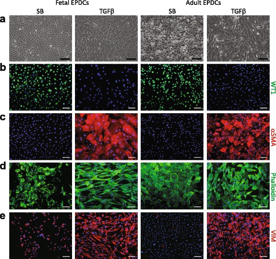
Fig. 3.

Human fetal and adult EPDCs undergo EMT upon TGFβ stimulation. a Phase contrast microscopy showed that SB-treated cells displayed a characteristic cobblestone morphology, while upon TGFβ stimulation both fetal and adult EPDCs underwent EMT. This was evident by transformation into a spindle-shaped and elongated morphology. Change in morphology was accompanied by (b) a decrease in nuclear WT1 and (c) an increase in αSMA. Upon TGFβ stimulation, (d) phalloidin visualized the increase in actin filaments and (e) VIM staining showed an organized network of intermediate filaments (scale bars: 100 μm). EPDCs epicardial-derived cells, SB medium containing SB431542, TGFβ transforming growth factor beta
