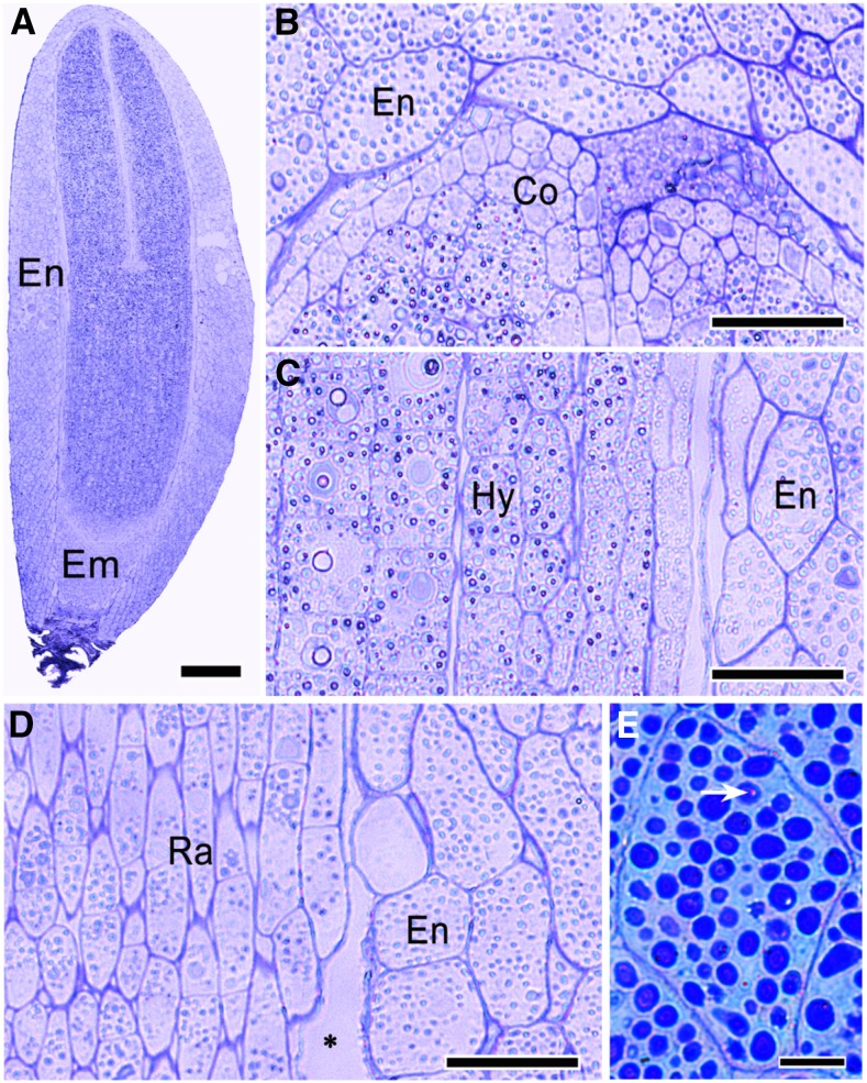
Figure 2.
Light micrographs of C. lanceolata seed sections. A, Semithin section stained with toluidine blue O indicates that the embryo is fully developed in a freshly matured seed. B to D, There is considerable variation in the morphology of cells in different parts of the embryo: (B) cotyledon, (C) hypocotyl, and (D) radicle. E, Protein bodies of an endosperm cell stained blue by Coomassie Brilliant Blue R. The asterisk in D indicates that there is a visible gap between the embryo and the endosperm in the radicle region. The arrow in E indicates the presence of polysaccharide in the cells stained by periodic acid-Schiff’s reagent. Co, Cotyledon; Em, embryo; En, endosperm; Hy, hypocotyl; Ra, radicle. Bars = 200 μm in A, 25 µm in B to D, and 10 μm in E.

