Figure 3.
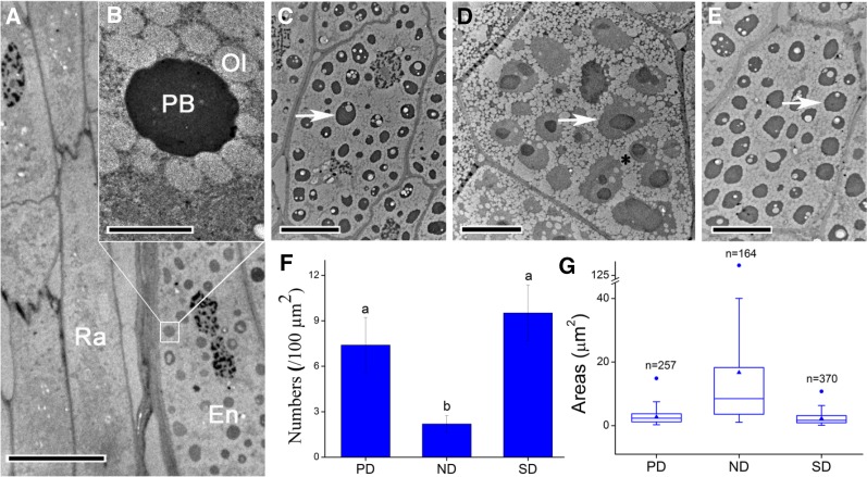
Electron micrographs of ultra-thin sections of the radicle region. A, Radicle cells and neighboring endosperm of a freshly matured seed. The endosperm cells contain many protein bodies, but the radicle cells do not. B, A higher magnification image of the region of interest in A, showing that protein bodies are surrounded by small oleosomes. C to E, Endosperm cells close to radicle cells in seeds with various dormancy statuses: (C) primary dormant, (D) nondormant, and (E) secondary dormant seeds. F and G, There is a notable change in number and size of protein bodies in the endosperm cells during dormancy release and induction: (F) number and (G) size. Numbers and sizes of protein bodies were calculated in 10 cells each of PD, ND, and SD seeds. There were 257, 164, and 370 protein bodies in the 10 cells of PD, ND, and SD seeds, respectively. The same lowercase letters in F and G indicate no significant difference (P > 0.05) between the values. Values in F are means ± sd; solid triangles indicate mean values, and solid circles indicate the maximum values in G. En, Endosperm; Ol, oleosome; PB, protein body; Ra, radicle. Arrows in C to E indicate PBs. The asterisk in D indicates that there was a coalescence of smaller oleosomes into a larger one. Bars = 10 μm in A and C to E and 2 μm in B.

