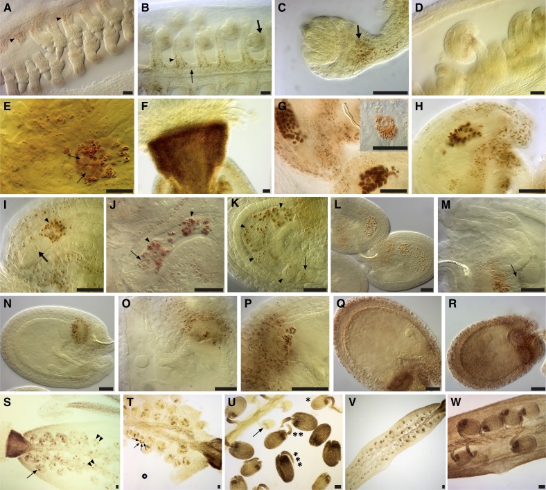
Figure 2.
Starch turnover during ovule and early embryo and silique development. A to C, During meiosis. The megaspore mother cell (A) and functional megaspore stages (B and C) are illustrated. Starch accumulated in the placenta (thin arrow), the funiculus (arrowheads), and the chalazal region below the nucellus (thick arrows). D, Division and elongation of the embryo sac, showing an extension of starch deposits to the integuments. E, Starch starts to build up in the central cell when the two polar nuclei lie next to each other (arrows). F, Strong starch accumulation in the parenchymatic cells of the style at anthesis. G and H, Ovules at anthesis (G) and during fertilization (H), showing more conspicuous starch accumulation in the central cell (surrounding the secondary endosperm nucleus; inset in G), the distal portion of the funiculus, the micropylar portion of the integuments, and the chalazal proliferating tissue. I to L, Starch accumulation is maintained in the dividing zygote (arrows) and endosperm (arrowheads: uninuclear in I, binuclear in J, four- or eight-nuclear stage in K and L). Note the trace of a discharged pollen tube in I (thick arrow). M to P, Starch disappears from all tissues but the chalazal proliferating tissue. Note the crushed nucellar cells (arrow) between the chalazal cyst of the endosperm and the starch-accumulating proliferating tissue in M, the pronounced ring-like shape of starch accumulation in N, the depletion of starch at the micropylar part of the seed in O, and the cells forming an inverted cup-like shape that are filled with starch and in close contact with the chalazal cyst in P. Q and R, A new wave of starch amylogenesis occurs at the octant embryo stage (Q) and is more conspicuous at the dermatogen stage (R); the endosperm is starchless. S to U, Low-magnification views of different postfertilization stages that are easily recognizable by their starch accumulation pattern and/or intensity. Uninuclear (arrows) and binuclear (arrowheads) endosperm stages are shown in S; four-nuclear endosperm stages (arrows) are shown in T; and unfertilized starchless ovule (arrow), quadrant (asterisk), octant (double asterisk), and dermatogen embryo stages (triple asterisk) are shown in U. Note the persistence of starch in the style and the beginning of basipetal starch accumulation in the silique walls and the placenta-septum region in S. V, A low-magnification view of a silique illustrating the basipetal starch accumulation in the valves. W, A middle silique portion with octant and dermatogen embryos, where conspicuous starch accumulation occurs in the valves and the placenta-septum region. Bars = 20 μm.

