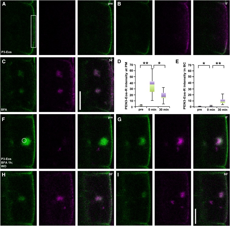
Figure 2.
PEN3-mEos2 endocytosis from and recycling to the outer lateral membrane. A to E, PEN3-mEos2 (P3-Eos) internalization from the outer lateral domain into BFA bodies after green-to-red photoconversion in the indicated ROI frame. A to C, PEN3-mEos2 treated with 50 μm BFA after photoconversion. Time points (A) prior to (pre) photoconversion, (B) after photoconversion (0′), and (C) after application of 50 μm BFA (34′) are indicated. D and E, Quantitative and statistical analysis of photoconverted PEN3-mEos2 (PEN3-Eos-R) intensity at (D) the PM or (E) in BFA compartments (BC). Box-and-whiskers plots are displayed for n = 10 cells prior to photoconversion (pre), directly after photoconversion (0 min), and 30 min after photoconversion. Whiskers indicate maximum and minimum values of the population. Violet boxes, 25% of values above median. Green boxes, 25% of values below median. Data are derived from CLSM images such as in A to C. Images at 30 min were acquired between 29 and 34 min after BFA application. Statistical differences were determined by two-tailed, type 1 paired t test with n = 10 cells (from seven roots). D, **P = 0.000 pre versus 0 min; *P = 0.003 0 min versus 30 min. E, *P = 0.001 pre versus 0 min; **P = 0.000 0 min versus 30 min. F to I, PEN3-mEos2 recycling from a BFA body to the outer lateral PM after photoconversion. After 60 min of 50 μm BFA pretreatment followed by BFA washout, photoconversion was conducted between pre (F) and 0′ (G) after BFA washout. PEN3-mEos2 redistribution monitored at indicated time points after BFA washout. n = 17 cells (from 15 roots) were observed with similar results. Bars = 10 μm.

