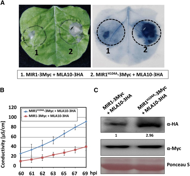
Figure 6.
MIR1 expression attenuates MLA10-triggered cell death in N. benthamiana. A, MIR1-3Myc or MIR1H104A-3Myc was coexpressed with MLA10-3HA in N. benthamiana. The photograph was taken at 70 h post infiltration (hpi; left), and then the leaf was stained with Trypan Blue solution (right). B, The electrolyte was measured using infiltrated leaf discs at the indicated time points. Error bars were calculated from three replicates per time point and construct. C, Immunoblot analysis of MLA10 protein level in N. benthamiana upon MIR1 and MLA10 coexpression. Total protein extract was obtained from N. benthamiana at 60 h post infiltration, and the experiment was done as described by Bai et al. (2012).

