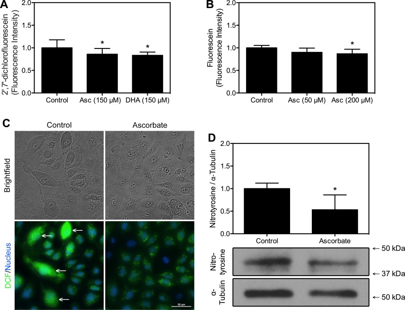
Fig. 2.
Ascorbate decreases oxidative stress markers in endothelial cells. A: HUVECs were exposed to 150 μM ascorbate (Asc), 150 μM DHA, or a media change for 1 h, then to 10 μM DCFH-DA in PBS for 1 h, and then rinsed and incubated in PBS for 1 h. Fluorescence intensity was measured and values were normalized relative to control; n = 24 wells per condition ± SD. B: BEnd.3 cells were exposed to 50 or 200 μM Asc for 30 min, to 20 μM dihydrofluorescin diacetate (DHF-DA) for 30 min, and then washed and incubated in Krebs-Ringer HEPES buffer (KRH). Fluorescence measures were taken at 0 and 36 min, and increase in fluorescence was quantified by subtracting initial from final values, which were then scaled relative to control; n = 7 wells per condition ± SD. C: HUVECs were treated as in A and DCF (green) and nuclei (Hoescht 33342; blue) were visualized as described in materials and methods. The reference bar shown measures 50 μm. White arrows point to discrete cells exhibiting high levels of fluorescence. Images are representative of 4 culture plates per condition. D: HUVECs were cultured for 5 days with 150 μM DHA-containing daily media changes and then lysed and subjected to Western blot analysis as described in materials and methods. The most prominent band formed at 42 kDa and was used for densitometry measures of nitrotyrosine, expressed relative to total α-tubulin; n = 4 lysates per condition. Molecular weight markers are indicated. Numerical results are shown as means ± SD. *P < 0.05, compared with the respective sample not treated with ascorbate or DHA.

