Fig. 6.
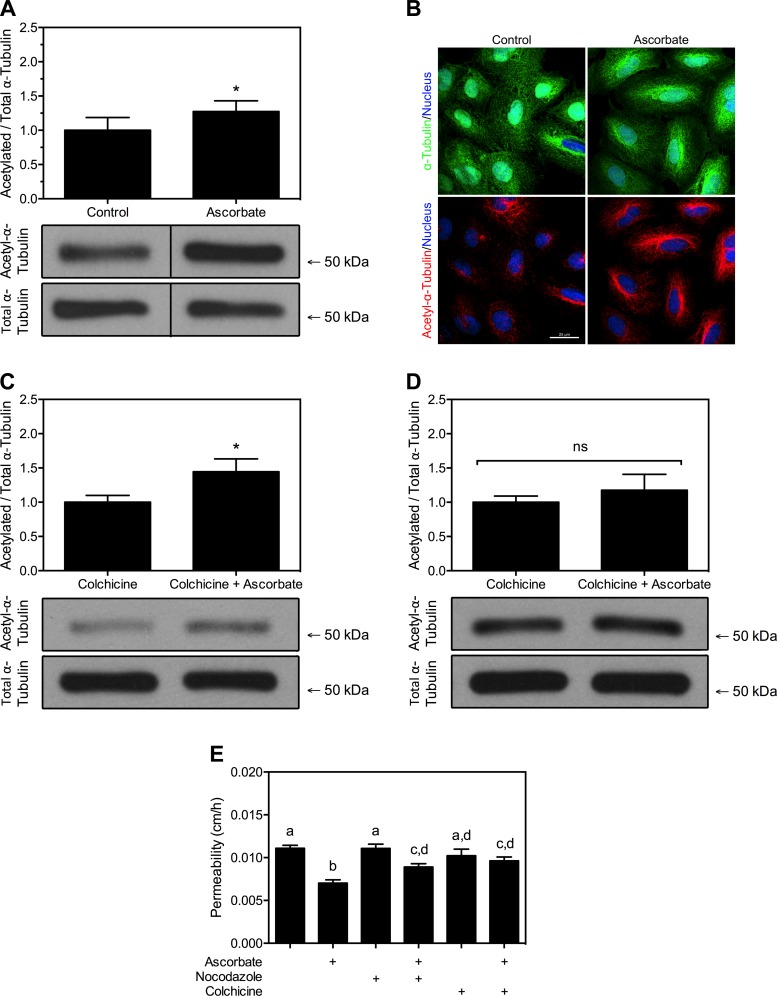
Ascorbate increases acetylated α-tubulin in HUVECs. A: HUVECs cultured with or without daily addition of 150 μM DHA for 5 days were lysed and probed for either total or acetylated α-tubulin via Western blot analysis, as described in materials and methods. B: HUVECs grown on coverslips were treated with DHA as described in A and then fixed and probed for total (green) and acetylated (red) α-tubulin with RedDot1 nuclear counterstain (blue). Images were captured with an Olympus FV1000 inverted confocal microscope at ×40 and are representative of at least 10 fields per condition. C: 150 μM DHA was added to HUVECs for 60 min and then 2.5 μM colchicine was added for 30 min. Western blot was performed as in A. D: 2.5 μM colchicine was added to HUVECs for 30 min and then 150 μM DHA was added for 60 min. Western blot was performed as in A. All Western blots had n = 4–5 samples per condition and are expressed as means ± SD. *P < 0.05; ns, no significance. Molecular mass markers are indicated. E: postconfluent HUVECs on Transwell filters were treated for 60 min with ascorbate and then for 30 min with 10 μM nocodazole or 2.5 μM colchicine. Cells were then subjected to an inulin transfer assay; n = 10 per condition. Bars not having the same lowercase letters are different at P < 0.05.

