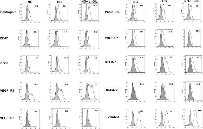
Fig. 10.
Expression of RPE cell markers. The RPE cells cultured in different glucose conditions were examined for expression of bestrophin, CD47, CD36, VEGF-R1, VEGF-R2, PDGF-Rα, PDGF-Rβ, ICAM-1, ICAM-2, and VCAM-1 by FACS analysis. The shaded area shows control IgG staining. Please note the increased level of bestrophin, CD47, CD36, and VEGF-R1 in high glucose and osmotic stress conditions, whereas increased ICAM-1, ICAM-2, and PDGF-Rβ expression levels only occurred in high glucose compared with control. The expression of VEGF-R2 and PDGF-Rα was low and did not change in different glucose conditions. In contrast, the level of VCAM-1 was significantly decreased in high glucose and osmotic stress. A decrease in PDGF-Rβ level was also noted in osmotic stress. The mean fluorescence intensities are shown for each sample. These experiments were repeated with two different isolations of RPE cells with similar results.

