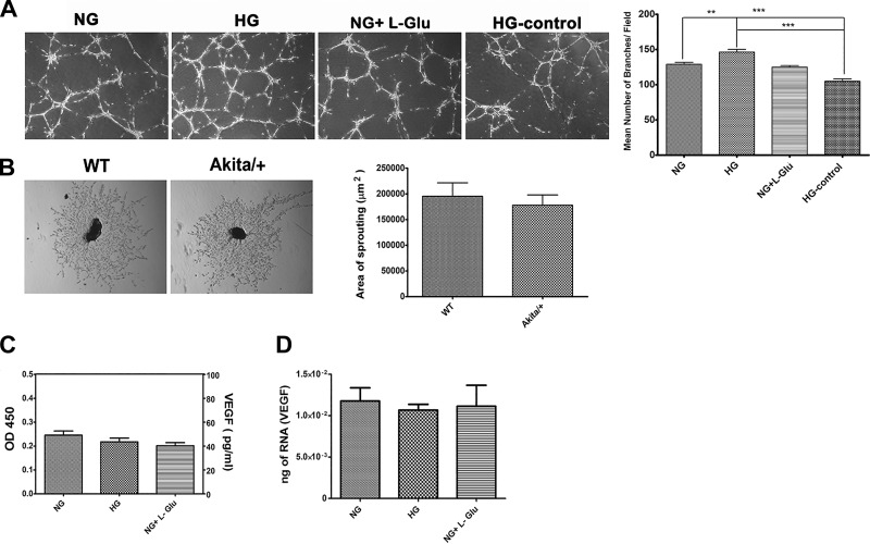
Fig. 12.
The capillary morphogenesis of choroidal EC and sprouting of RPE-choroidal explants. A: the capillary morphogenesis of choroidal EC incubated with conditioned medium prepared from RPE cells in different glucose conditions. Unconditioned high glucose medium was used as a control. Please note a significant increase in branching morphogenesis of choroidal EC incubated with conditioned medium prepared from RPE cells cultured in high glucose (**P < 0.01 and ***P < 0.001, n = 3). B: sprouting of choroid-RPE tissues prepared from 3-mo-old wild-type and Akita/+ mice. Images shown here represent results obtained from five different mice with the desired genotype (×40). Please note no significant difference was observed in the degree of choroidal sprouting in Akita/+ mice compared with wild-type mice (P > 0.05, n = 5). C: the level of secreted VEGF protein was determined in conditioned medium collected from RPE cells in different glucose conditions by using an ELISA. Please note there was no significant difference in the amount of VEGF produced by RPE cells in different glucose conditions (P > 0.05, n = 3). D: the level of VEGF mRNA was determined by qPCR using RNA prepared from RPE cells in different glucose conditions. Please note there was no significant difference in the amount of VEGF mRNA detected in RPE cells in different glucose conditions (P > 0.05, n = 3).

