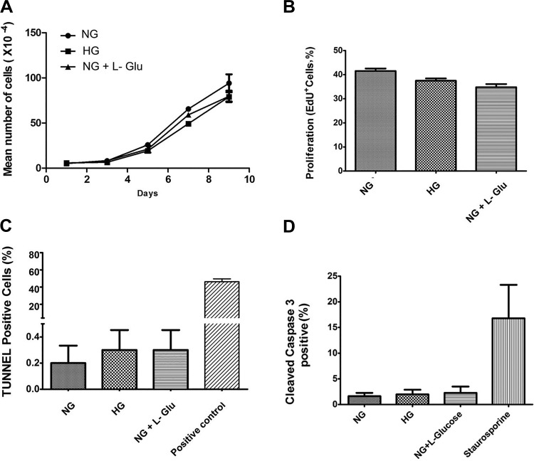
Fig. 2.
High glucose conditions minimally affected the proliferation and apoptosis of RPE cells. A: the rate of RPE cell proliferation incubated under various glucose conditions was determined by counting the number of cells as detailed in materials and methods. High glucose conditions did not affect the proliferation rate of RPE cells compared with controls. B: the rate of DNA synthesis in RPE cells under various glucose conditions was determined by EdU labeling. No significant changes in the rate of DNA synthesis was observed in RPE cells under high glucose conditions compared with controls. C: the rate of apoptosis in RPE cells cultured in different glucose conditions was determined by TdT-dUTP terminal nick-end labeling (TUNNEL) assay. Please note no significant differences were observed in the apoptosis of RPE cells in different glucose conditions (P > 0.05, n = 3). D: similar results were observed by determining the percentage of cells that were positive for cleaved caspase-3 in different glucose conditions (P > 0.05, n = 3).

