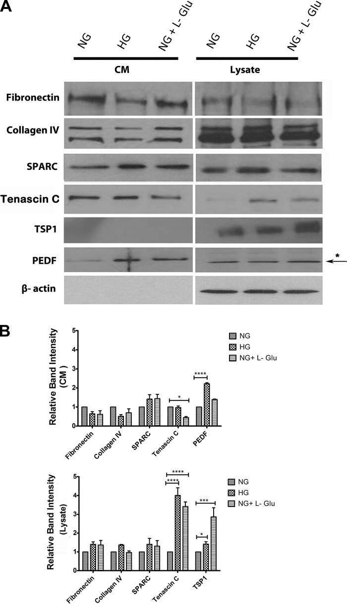
Fig. 5.
Altered expression of ECM proteins in RPE cells. A: RPE cells were plated on gelatin-coated 60-mm dishes in different glucose conditions for 5 days and then were incubated for 48 h in serum-free growth medium in different glucose conditions. The conditioned medium (CM) and cell lysates were prepared for Western blot analysis of different ECM proteins as described in materials and methods. The expression of fibronectin, collagen IV, tenascin C, TSP1, PEDF, and SPARC were determined using specific antibodies. The β-actin was used as a loading control for cell lysates. Please note a significant increase in the level of tenascin C and TSP1 in cell lysates in high glucose and osmotic stress compared with normal glucose. B: the quantitative assessment of the data (*P < 0.05, ***P < 0.001, and ****P < 0.0001, n = 3). Only the level of PEDF was significantly increased in conditioned medium of RPE cells in high glucose. We also examined the levels of periostin, opticin, and osteopontin and there were no significant changes (not shown).

