Fig. 6.
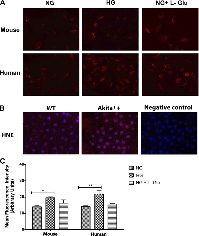
Increased oxidative stress in RPE cells in high glucose. A: the level of oxidative stress in mouse and human RPE cells was assessed by dihydroethidium staining. A significant increase in the level of ROS was detected in high glucose in both mouse and human RPE cells compared with normal glucose or osmotic stress. B: Wholemount staining of RPE-choroid tissues prepared from wild-type and Akita/+ mice with antibody to 4-hydroxy-2-nanonal (HNE). Please note increased HNE staining in RPE-choroid tissues from Akita/+ mice compared with wild-type mice. These experiments were repeated with two different isolations of RPE cells and eyes from five different mice with similar results. C: the quantitative assessment of data in A (*P < 0.05 and **P < 0.01, n = 3).

