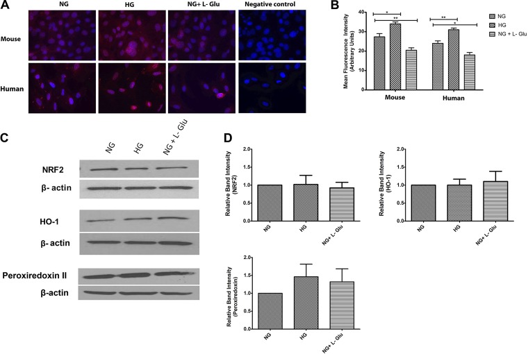
Fig. 8.
Cellular localization and expression of NRF2 and its downstream target genes Ho-1 and Prdx2. A: the localization of NRF2 was determined by immunofluorescence staining. Mouse and human RPE cells were plated on fibronectin-coated chamber slides in different glucose conditions and stained with specific antibodies as detailed in materials and methods. No staining was observed in the absence of primary antibody (negative control). Please note increased nuclear localization of NRF2 in both human and mouse RPE cells in high glucose. B: the quantitative assessment of the data (*P < 0.05 and **P < 0.01, n = 3). C: the expression of NRF2, HO-1, and peroxiredoxin II was determined by using specific antibodies. The β-actin was used as a loading control. D: the quantitative assessment of data (P > 0.05, n = 3).

