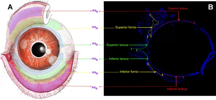
Fig. 1.
Demarcation of conjunctival regions. A: scheme of the stereotypical conjunctival epithelial architecture of adult mouse [6- to 8-wk-old mice (C57BL/6J, Harlan) of both sexes were used in all experiments]. The purple areas (red arrows) extending from the superior and inferior lid margins indicate the superior and inferior tarsus, the green curved ribbon (yellow arrows) adjacent to the bulbar conjunctiva indicates the superior and inferior fornix, and the half ring-shaped blue curve (green arrows) marks the superior and inferior bulbus. B: sagittal frozen section of mouse conjunctiva (n = 21 in 3 independent experiments). Cell nuclei were visualized with DAPI (blue). The superior and inferior conjunctiva formed a continuous envelope shape with each region marked and labeled with arrows. Cells and explants were obtained from the individual sections labeled in A and B. Scale bars, 500 μm.

