Figure 3.
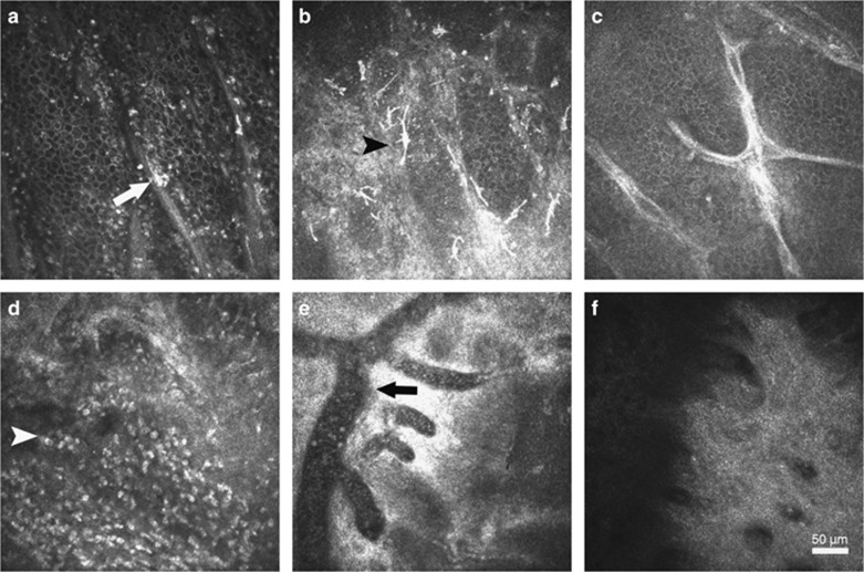
In vivo confocal microscopic images of limbus after ocular chemical injury. Typical image of the Palisades of Vogt (POV) was found in the eye with grade I injury (a), which was characterized by alternant epithelium–stromal cords with a bright fringe of hyperreflective basal epithelial cells. A few dendritic cells were visible (white arrow). More dendritic cells (black arrowhead) were found to infiltrate within the edematous epithelium–stromal cords in the eyes with grade II injury (b). The atrophic POV was identified in the eyes with grade III injury (c), which was characterized with the absence of hyperreflective basal epithelial cells around stromal cords. In the eyes with grade IV injury, numerous inflammatory cells (white arrowhead) instead of POV were found in the limbus (d), along with distinctly dilated vessels (e). At 3 months, the formation of acellular fibrous structure was found at limbus in the eyes with grade IV injury (f).

