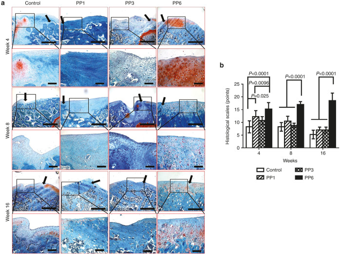
Figure 4.
Histological examination of articular cartilage (AC) repair with BMP4-expressing muscle-derived cells (MDCs). (a) Safranin-O staining of the defect areas at week 4, week 8, and week 16 post-transplantation; Black arrows indicate the edge of the defect areas; upper panel (Scale bar = 500 μm, original magnification × 40), lower panel (Scale bar = 200 μm, original magnification × 200). Control: fibrin glue only without cells. (b) Chronological changes of the histological grading score. n = 4, data was presented as mean ± SD.

