Figure 1.
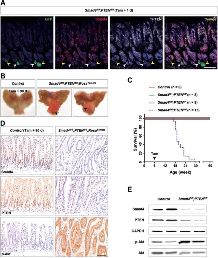
Deletion of Smad4 and PTEN in murine gastric Lgr5+ stem cells resulted in tumorigenesis. (A) Immunofluorescence (IF) analysis using Smad4 (red), PTEN (white) and GFP (green; for Lgr5+ cells) antibodies in Lgr5+ stem cells from Lgr5-CreERT2;Smad4fl/fl;PTENfl/fl (hereafter Smad4fl/fl;PTENfl/fl) antrum 1 day after tamoxifen (Tam) administration. White arrowheads indicated the wild-type Lgr5+ stem cells, and yellow arrowheads indicated the deficient Lgr5+ stem cells. (B) Gross anatomy of the representative stomach of control and Smad4fl/fl;PTENfl/fl;RosaTomato double-mutant mice 90 days after tamoxifen treatment; the latter shows macroscopic polyps (arrowheads). Of note, red polyps could be clearly seen without a microscope. (C) Kaplan-Meier survival curve of Smad4fl/fl;PTENfl/fl compound mutant mice (blue dotted line; n = 15) compared with control mice (red solid line; n = 8; P < 0.0001 by log-rank test), Smad4fl/+;PTENfl/fl mice (green solid line; n = 8; P < 0.0001) or Smad4fl/fl;PTENfl/+ mice (purple solid line; n = 8; P < 0.0001). (D) IHC analysis of Smad4, PTEN and p-Akt confirmed complete loss of Smad4 and PTEN and activation of Akt. (E) Western blot analysis of Smad4, PTEN and p-Akt in the control antral epithelium and Smad4fl/fl;PTENfl/fl polyps 90 days post induction. Scale bar, 50 μm.

